A Blood Smear Should Contain Which of the Following:
Normal synovial fluid should contain no erythrocytes and moderate numbers of nucleated cells. The smear provides this information.

I Love All These Cells But My Favorite Is The Plasma Cell Hematology Medical Laboratory Science Medical Lab Technician
Fixation of smear is because of precipitation of proteins by alcohol which prevent washing off of the film.
:max_bytes(150000):strip_icc()/iStock-470812556-5c74be3ec9e77c0001d19bd8.jpg)
. A good quality smear has the following features. Place the first step at the top 1. Cells of normal size and hemoglobin content color are termed normocyticand normochromic.
Blood drop should be of proper size. When latex or vinyl strap tourniquets become soiled with blood it is best to. Red blood cells RBCs erythrocytes carry oxygen to tissues.
The solution normally used to clean the site before routine venipuncture is. Increased red cell destruction. Let the blood dry.
All parts of the smear should be examined however the monolayer is the area where the cells are examined in close detail and differential cell counts performed. Nucleated red cells in the peripheral circulation. The blood smear allows for the evaluation of these cells.
Angle should be maintained at 45. The number and kinds of white blood cells differential or percentage of each type of cell The number and kinds of abnormally shaped blood cells. It is pink or red in colour.
Microcytic hypochromic red cells are most often associated with impaired. Red blood cells have a central concavity that. Components of a blood smear.
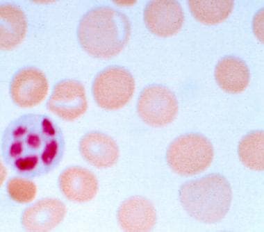
White blood cells WBCs leukocytes help fight infections or participate in immune responses. A blood smear is a sample of blood thats tested on a specially treated slide. Most blood collection tubes contain an additive that either accelerates clotting of the blood clot activator or prevents the blood from clotting anticoagulant.
A blood smear is considered normal when your blood contains a sufficient number of cells and the cells have a normal appearance. Wright stain which contains methylene blue and stains acidic cellular elements cytoplasmic RNA and nuclear DNA variations of blue and Giemsa stain which contains eosin and stains basic cellular elements hemoglobin and some cytoplasmic. The blood smear test shows a sample of blood components including platelets leukocytes white blood cells and erythrocytes red blood cells that are present in plasma the fluid part of blood.
Platelets thrombocytes small cell fragments that are vital to proper blood clotting. A well made blood smear consists of several areas. Examination of the peripheral blood smear should be considered along with review of the results of peripheral blood counts and red blood cell indices an essential component of the initial evaluation of all patients with hematologic disorders.
A blood smear test is said to be normal when the sample of blood contains an optimum number of cells and the cells have a normal appearance. Peripheral Blood Smear Test Results Fully Explained. Methylene blue is a basic dye that stains acidic structures like nucleus or granules of basophils.
The focus will be on the three primary types of cells that can be found within the blood. Spreaders edges should be smooth and it should be smaller than the slide on which smear is being made. Rank the steps below into the correct sequence necessary to prepare a blood smear.
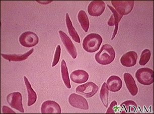
A blood smear is considered abnormal when theres an abnormality. Obtain a drop of blood and place it away from the center onto a slide 2. The presence of schistocytes on the peripheral blood smear is commonly associated with.
GENERAL PRINCIPLES OF REVIEWING A PERIPHERAL BLOOD SMEAR. This edge should have a fine feathery appearance. Peripheral blood smears are routinely stained with.
Small red cells are microcytesdiameter less than 6 μm. The examination of blood films stained with Wrights stain frequently provides important clues in the diagnosis of anemias and various. A well-developed feathered edge.
Red blood cells RBCs white blood cells WBCs and platelets. Drops of blood are smeared across a microscope slide to be examined by an expert in a lab. This should take up about 23 of the entire smear and should blend smoothly into the monolayer area.
For a blood smear test a laboratory professional examines the slide under a microscope and looks at the size shape and number of different types of blood cells. Leukemia anemia malaria and numerous other blood conditions can be identified with a. What may be a sign of accelerated bone marrow erythropoiesis.
It is blue in colour. The list below lists the most commonly used blood collection tubes their additives and uses in laboratory. Larger than normal erythrocytes are macrocytesdiameter greater than 9 μm.
Red blood cells which carry oxygen from your lungs to the rest of your body. CBC Part 2 WBC differential blood morphology CBC Part 3 RBC morphology platelet estimate CBC Part 4 Post-test The review of hematopoiesis and blood cell morphology ie Hematology Atlas located on the LSUHSC server is recommended as a. Take a clean slide and slide it at a 45 degree angle across the first slide to distribute the drop of blood evenly on the first slide.
A blood smear is basic test that is used to determine diagnostically if there are any abnormalities within the blood. The results of the peripheral smear are considered abnormal when there is an abnormality in the shape size or number of cells in the blood sample. The feathered edge the monolayer and the body and base of the smear.
Pressure applied should be proper. They are anucleate non-granulated eosinophilic cells that are uniform in shape biconcave discs and size 72 microns. Or the blood may be examined by an automated machine.
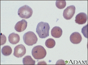
Acetone free methyl alcohol is a fixative for smear. Erythrocytes or red blood cells are by far the predominant cell type in the blood smear. And those with central pallor greater than 50 of the diameter are hypochromic.
Blood Smear Information Mount Sinai New York

Making A Great Blood Smear Sonopath
Which Peripheral Blood Smear Findings Indicate Pernicious Anemia

Blood Smear Shotgun Histology Youtube

Peripheral Blood Smear Using The Sample Collected In The Er Of The Download Scientific Diagram

Blood Smear Iron Deficiency Uptodate

Trypanosoma Classification Characteristics Life Cycle Microscopy Medical Laboratory Science Plasma Membrane Medical Laboratory

Making A Great Blood Smear Sonopath
Blood Smear Information Mount Sinai New York

Making A Great Blood Smear Sonopath

How To Identify The Type Of Malaria On A Blood Smear Medmastery

Pathophysiology Peripheral Blood Smears Flashcards Quizlet

Pathophysiology Peripheral Blood Smears Flashcards Quizlet
2

Clinical Case Challenge What Can You Learn From A Peripheral Blood Smear News Center At Cummings School Of Veterinary Medicine At Tufts University
Blood Cytology
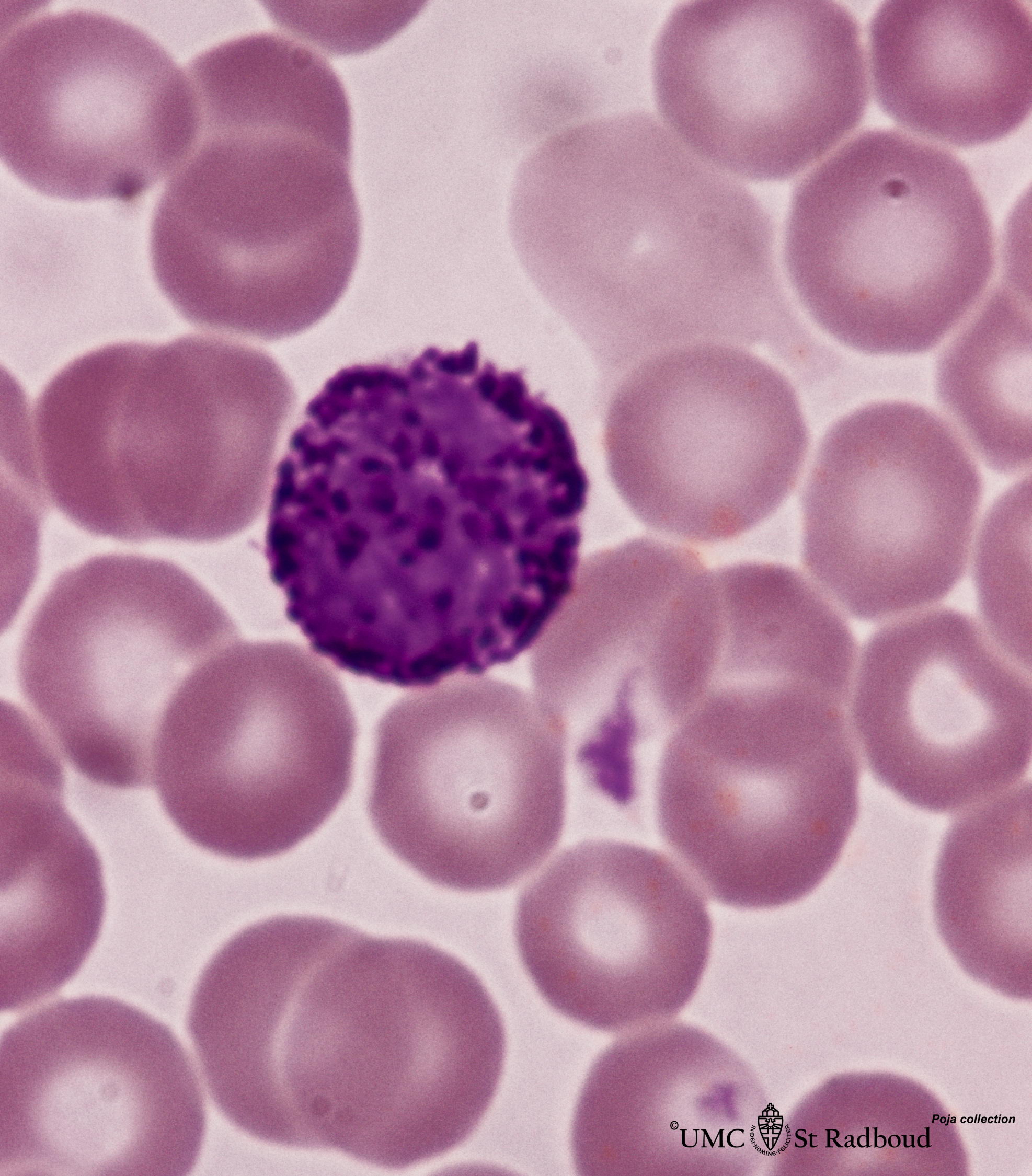
Basophilic Granulocyte In Peripheral Blood Smear Human Eccles Health Sciences Library J Willard Marriott Digital Library
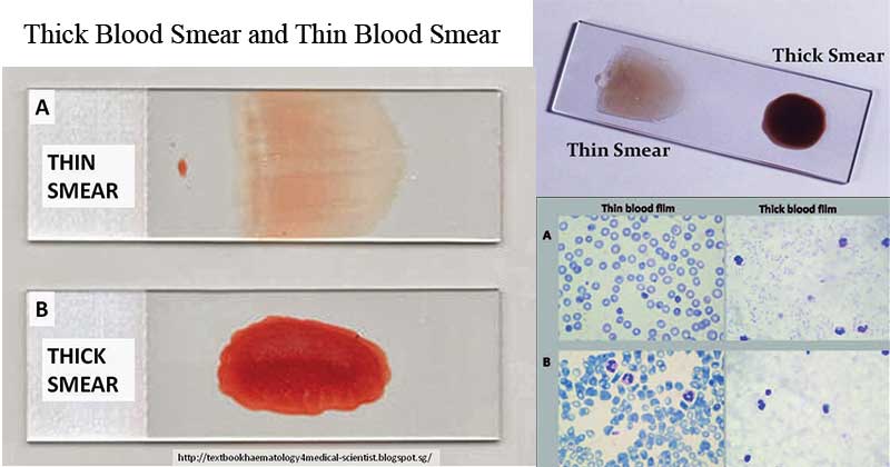
Thick Blood Smear And Thin Blood Smear
Blood Smear Uses Side Effects Procedure Results


Comments
Post a Comment